|

|
>>>>>
Special
Products
– Micromed
|
|
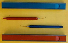 |
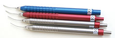
Cannulas
Buratto-cruse
The cannulas of Buratto developed
by MICROMED are cannulae for the
suction of residues bimanual cortical
after facomulsificazione of the
nucleus. They are used through the
tunnel corneal and allow to reach
all points of the bag for complete
removal.
Buratto's cannules by MICROMED are
developed for bimanual infusion
of cortical remainings after facoemulsification
nucleus. Used through corneal tunnel,
they allow operators to reach all
parts for a complete residual removal. |
|
|
|
|
|
|
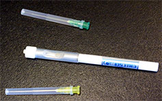
Tubes
of double tumbler
Cannulas in both hands for
the extraction of residual cortical
surgery FACO and ECCE.
allow easy and convenient cleaning of
the capsule even in the most hidden. |
|
|
|
|
|
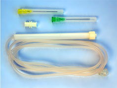
Suction
Hand Pieces
Suction handpiece and Minimally
Invasive Vitrectomy with silicone button
for reflux (aspiration spontaneous type
Charles)
|
|
|
|
|
|
Handful
of aspirazioneper vitrectomy and minimally
invasive
(Active
suction with connection to the vacuum
line)
The final infusion cannulas and / or
extraction of silicone oil are defined
court and specifically have measures:
- 20G (0.9mm) x 8mm
- Teflon 18mm 60 ° cutting
The cannulas intended for handpieces,
the perfluorocarbon liquid and protected
areas are defined long and specifically
measures are:
•
20G (0.9mm) x 31.3mm
•
23G (0.6 mm) x 31.3mm
•
25G (0.5 mm) x 31.3mm
•
20G x 31.3mm protected (+ 4mm silicone
tube protruding)
|
|
|
|
 |
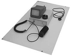
Surgical
simulator for training MMD-150
A support device for animals bulbs
The kit contains:
•
Model of human head (actual size)
•
Bowl pick-liquid
•
Element portabulbi
•
Transformer
In particular element portabulbi
is as follows:
•
Upper ring nut for fixing the eyeball
teflon white (we recommend using an
eye pig)
•
5 interchangeable rings in aluminum
anodized in black (depending on the
size of the eye used)
•
Housing of the eyeball, in white Teflon
•
High brightness LED (lights up the back
of the eye)
•
Steel ring (in the desired direction
to guide the eye)
•
Body and base of the steel portabulbo
•
Screw blocker (so you can place the
eyeball to the desired height)
•
Electrical cable (transformer and current)
|
|
|
 |
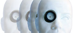
MMD-110
Videopupillometro of Cheesecloth
The videopupillometro IR is a device
capable of observing the eyes in conditions
of absence of actinic light. It is based
on a high-resolution CCD TV system with
very strong sensitivity in the range
of 900 nm. Has optics capable of projecting
an enlarged image of the pupil on the
sensor, which covers up to 70% of the
surface (fully dilated pupil).
A light-tight bellows allows measurements
to be made with ambient lighting and
not in the dark. E 'can then with generators
(IR emitting solid state) produce highly
contrasted lighting board pupil and
a reflection on the cornea net to ensure
that the position of the iris is perfectly
parallel to the CCD.
The detected signal from the camera
is presented for viewing on a monitor
and combined with a grigla graph that
gives the objective reference of pupil
diameter. At the time of acquisition
the image is stored and then printed
(set to the grating reference) to provide
documentation of the exact pupillary
diameter is that both the correta focus
dell'orletto. Beyond complete visibility
in the dark pupil then, for an exact
measurement of the diameter is necessary
that the eye is in position exactly
perpendicular to the iris and the rib
is perfectly in focus.
With this adapter you can view
images of viodeopupillometro directly
on the computer monitor, with obvious
advantages in terms of documentation
and measurement and printing. The data
acquisition is done by push butt
|
|
|
|
|
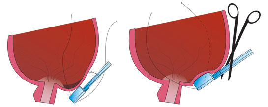
Macular
button the ACILITIES for scleral buckling
According Sons - Ripandelli
The technique
Plant
silicone for use in high myopic patients
during surgery for the posterior detachment
of the retina.
Using this system it is possible to
perform a particular surgical technique
in order to obtain the complete riadagiamento
retinal raised with greater safety and
efficacy than those generated with the
traditional techniques with silicone
implants to rear block until now in
use.
Description
The
plant looks like a block of parallelepiped
shape with a thickness of 4 mm having
surfaces that may vary between 5 x 5
mm and 8 x 8 mm.
On the edge orthogonal to the thickness
of the block and 'applied also to form
a handle parallelepiped of the size
of 2 x 2 x 10 mm which will be used
to adjust the proper implant placement
and then severed and removed only at
the end of the surgical procedure.
and 'printed in silicone rubber biocompatible
certified for long residence times in
contact with the eye and the hardness
of at least 90 shore.
edges of the box are beveled to avoid
trauma irritation scleral surfaces.
|
Application
Before
applying the two quotes arm opposite to
the handle with two-wire non-absorbable
and turning the eye upward to expose the
apical point of the equatorial sclera corresponding
to the macular region, and then slide the
lock by pushing it with the handle (the
periphery) until will not be positioned
on the macula.
Subsequently suture the handle (near to
its attack with the block) to the temporal
edge of the staphyloma along the meridian
that intersects the foveal region.
Apply to this point two sutures to block
the terminal part of the handle (the one
facing the block) firmly in place.
Thus check ophthalmoscopic if the traction
of the two wires the indentation of the
system falls in the desired zone; otherwise
repeat positioning.
When the indentation is centered at the
posterior detachment of the retina connect
the implant head, by means of wires not
absorbable, performing two sutures in place
at the level of the nasal sclera device,
safer, in correspondence of the meridians
of the 6 o'clock and 12.
Once assured the block and checked that
indenti properly is possible to fix the
sutures and cut the handle, concluding thus
the intervention.
This procedure minimizes the risks associated
with the passage of the needles and sutures
through the thin scleral wall of these patients.
Conclusions
The
procedure described allows to avoid excessive
surgical risks due to simplification of
the surgical maneuvers which have a lower
technical difficulty; at the same time,
through the wires that support the apex
of the block and that will be fixed in the
areas scleral multiple devices, ensures
a greater chance of a correct anatomical
location of the plant with a superior gradation
of its effect indentante.
During the operation is in fact possible
to direct control by the surgeon about the
location of the body of the button compared
to the area to indent and efficacy of '
indentation sought.
Such adjustments are possible even after
adjusting sutures depended on the proper
placement of the implant silicone in the
sense of greater invagination and relaxation
during the checks carried out in the postoperative
period.
For Further Details Visit : www.micormed.it
|
|
|
|
|
|
|
|